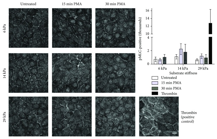
Figure 5.
PMA treatment did not increase pMLC localization to actin stress fibers in cells on varied stiffness substrates. PAEC monolayers on 6, 14, or 29 kPa gels were treated with 1 μM PMA for 15 or 30 minutes, prior to fixation and pMLC immunofluorescent labeling. For the positive control, cells on a 29 kPa gel were treated with 10 U/mL thrombin for 30 minutes. Images are maximum intensity projections from 60x confocal z-stacks. Scale bar is 25 μm. pMLC-positive pixels were quantified using the custom MATLAB code. Stiffness and PMA treatment were not significant by n-way ANOVA.

