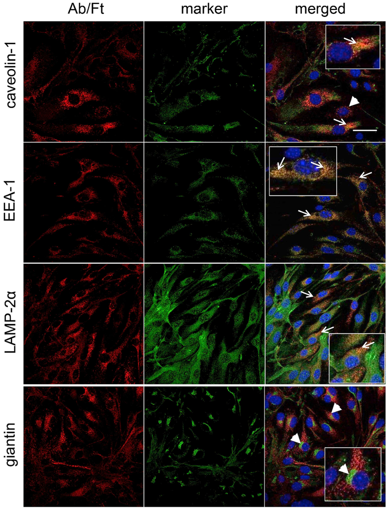
Fig. 7. Co-localization of Plvap-directed Ab/(Ft/SOD) with cellular organelles.
Cells were incubated with nanoparticles for 1 h at 37 °C, washed, fixed, permeabilized and stained for the ferritin nanoparticles with labeled secondary anti-rat IgG (red) and the following markers (all are in green): caveolin-1 (caveolae), EEA-1 (endosomes), LAMP-2α (lysosomes), giantin (Golgi). Nuclei were stained with DAPI (blue). Representative images correspond to 0.3-μm slice. Yellow-colored area indicates colocalization of nanoparticles and organelle. Arrows show co-localization of nanoparticles with organelle marker, arrow heads - no co-localization. Zoomed views are inserted in merged panels. Confocal microscopy. (For interpretation of the references to color in this figure legend, the reader is referred to the Web version of this article.)

