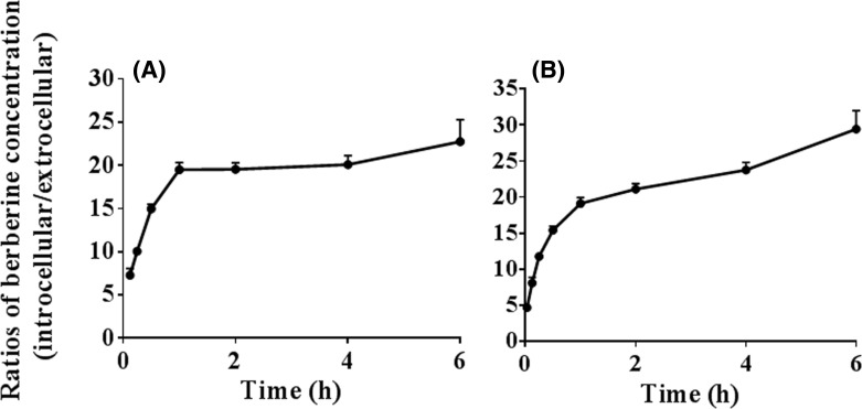
Figure 1. Accumulation of berberine in the HepG2 cells (Mean ± S.D., n=3).
The cells were incubated with berbrine [1 (A) or 10 μM (B)] at 37°C for different times [0.033 (only for 10 μM), 0.125, 0.25, 0.5, 1, 2, 4, or 6 h]. The incubation medium was collected at the designated time points. The cells were then washed three times with chilled PBS and harvested by trypsinisation. The cells were stored in 0.5 ml chilled water and were broken by repeated freezing and thawing. The concentration of berberine in the incubation medium and the cell homogenate was quantified using the LC-MS/MS method. The ratios of the intracellular to extracellular (i.e., the cell homogenate to incubation medium) concentration of berberine were calculated to reflex the cellular accumulation of berberine.

