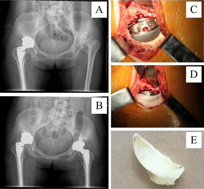
Figure 1A-E.

Acetabular reconstruction and methods of determining the area of uncovered acetabular cup in vivo. A preoperative (A) and postoperative (B) anteroposterior pelvic radiograph. (C). After acetabular reconstruction during surgery, the uncovered surface of cup was shown. (D). Bone wax pressed on the uncovered surface of the acetabular cup. (E). Bone wax model representing the uncovered surface was removed from the cup.
