Figure 2A-D.

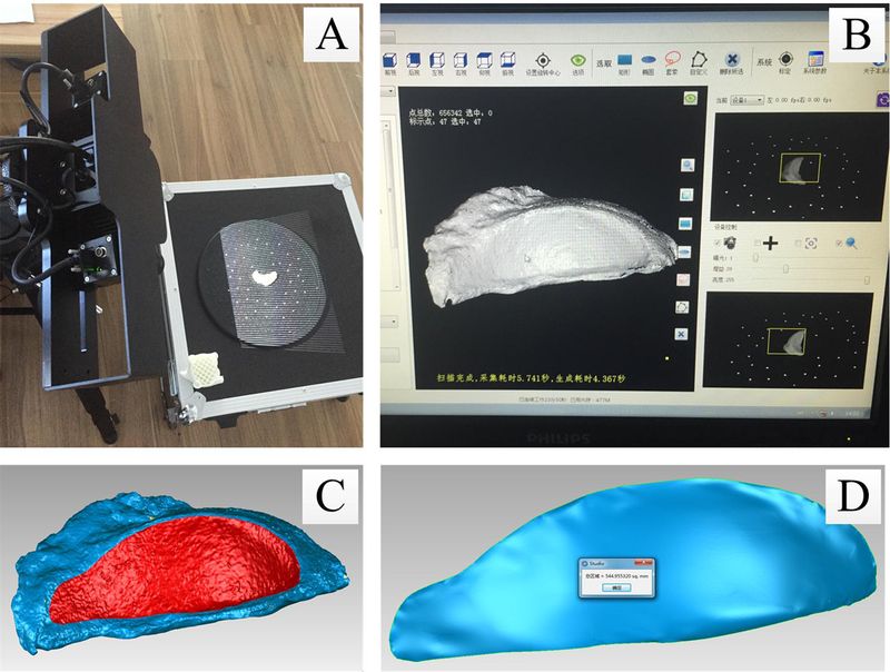
3D scan and processing of the bone wax model representing the uncovered surface of the acetabular cup. (A). 3D scanning. (B). Point cloud data processing. (C). Surface identification. (D). Area calculation.

3D scan and processing of the bone wax model representing the uncovered surface of the acetabular cup. (A). 3D scanning. (B). Point cloud data processing. (C). Surface identification. (D). Area calculation.