Figure 1.
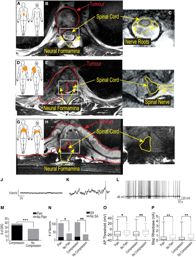
DRG neurons from dermatomes with radicular/neuropathic pain show ectopic spontaneous activity and hyperexcitability. Pain diagrams and MRI spinal images for three categories of patients are shown in A–I. The orange shaded area in A, D and G indicate where patients marked the location of their pain. This was either localized to the spine without signs of radicular/neuropathic pain (axial pain only, A); showed radiation only to one side (unilateral radicular/neuropathic pain, D); or pain that radiated to both sides of the body (bilateral radicular/neuropathic pain). The large MRI scan in B shows that patients with axial pain often only had tumours (outlined in red) that did not compress the nerve roots or spinal cord. Patients with unilateral neuropathic pain (E) typically had tumours that compressed one or more nerve roots on one side and part of the spinal cord. Patients with bilateral neuropathic pain typically had compression of one or more roots on both sides and the spinal cord (H). The area in B, E and H outlined in white are magnified in C, F, and I to show the spinal cord and nerve roots better (outlined in yellow). A representative recording of the resting membrane potential with an expanded time base for a cell without spontaneous activity is shown in J while a similar recording for a cell with spontaneous activity is shown in K to illustrate the spontaneous depolarizing fluctuations (DSFs) in membrane potential that occurred in these cells. A single action potential is shown at the right of this trace occurring atop one of the larger of these DSFs. The representative trace shown in L illustrates the irregular pattern of action potentials typically seen in cells with spontaneous activity. The bar graphs in M show that radiological evidence of nerve compression was strongly associated with signs of radicular/neuropathic pain; while in N the bar graphs show the relationship of radicular/neuropathic pain and nerve compression with spontaneous activity (SA). The box and whisker plots in O and P show that DRG neurons from a dermatome with pain and/or nerve compression had a more depolarized spike threshold potential and lower rheobase, respectively.

