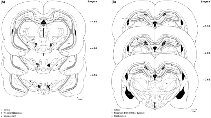
Figure 1.
(A) Coronal schematic sections show the microinjection sites in the ventral tegmentum areas (○ Vehicle (DMSO); ● Orexin A microinjection; ▲ Misplacement). D3V: Dorsal 3rd ventricle; cc: Corpus callosum; fr: Fasciculus retroflexus; str: superior thalamic radiation; PC: Paracentral thalamic nucleus; 3V: 3rd ventricle; ml: medial lemniscus; ML: medial mammillary nu, lateral; SuM: Supra mamillary; PBP: Parabrachial pigmented nucleus; SNR: Substantia nigra, reticular part. (B) Coronal schematic sections show the microinjection sites in the hippocampus CA1 (○ Vehicle (saline); ● SCH 23390 or Sulpiride microinjection; ▲ Misplacement). DG: Dentate gyrus; CA2: Field of CA2 of the hippocampus; CA3: Field of CA3 of the hippocampus; MoDG: Molecular layer dentate gyrus; D3V: Dorsal 3rd ventricle; cc, Corpus callosum; LV: Lateral ventricle; 3V: 3rd ventricle; Slu: Stratum lucidum. hyppocampus; Po: Post thalamic nuclear group; f: Fornix; mt: mammillothalamic tract

