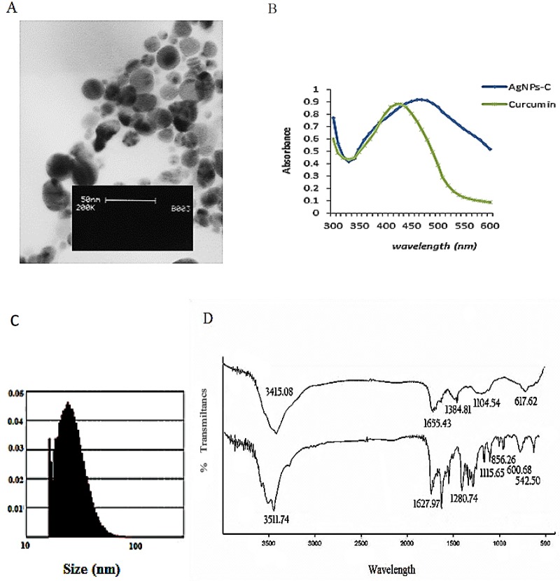
Figure 1.
(A) TEM image of AgNPs-C, (B) Uv- visible spectrom from solution contains AgNO3 and curcumin after passing 24 h,
(C) particle size disruption of AgNPs-C, (D) comparing FTIR spectra from AgNPs-C (A) and pure corcumin (B) theses spectra are very similar which indicated that curcumin coated the surface of silver nanoparticles.

