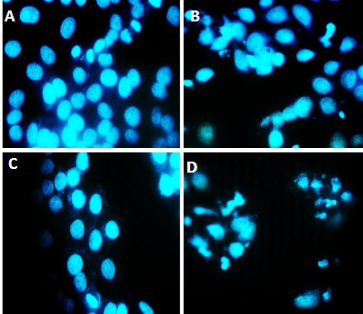
Figure 4.

DAPI nuclear staining, (A) Represents control group that did not receive any treatment and cell nuclei are intact, (B) Represents the group that treated with only silver nano particles in this group some nucleus of cells are fracture, (C) Cells treated with cisplatin. The most cell nuclei are intact, (D) Cells treated with cisplatin and silver nanoparticles together. In this group cell nucleus are fractures and showed chromatin condensation (arrow), the apoptotic features in synergistic groups was higher compared to groups that were treated with cisplatin or silver nanoparticles alone were used
