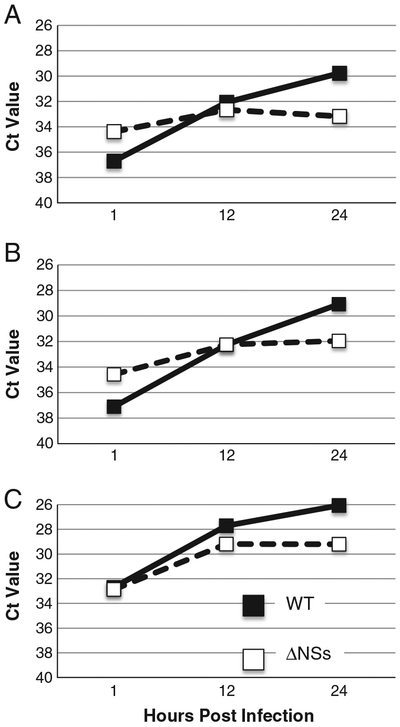
Fig. 3.
ΔNSs RVFV replicates to lower levels than wild-type RVFV in MDM. MDM were infected with WT or ΔNSs RVFV. RNA was purified from cells at various times post infection and analyzed by real time PCR. WT virus (black squares with solid lines) replicated to higher levels than the ΔNSs virus (white squares with dotted lines) on cells from the same donor. Data are presented as inverse Ct value at various times post infection. 3 different donors are represented in the figure. RNA from the 4th donor was not available for testing.

