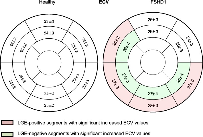
Fig. 6.
Assessment of myocardial fibrosis- Comparison of all patients with FSHD1 and healthy subjects with AHA segments showing ECV values in %. Significant differences between healthy suybjects and FSHD1 patients were found not only in LGE-positive segments (basal inferolateral: p < 0.01, basal inferior: p < 0.01, basal anteroseptal: p < 0.01, basal inferoseptal: p = 0.027), but also within the adjacent regions (medial inferolateral: p = 0.048, medial inferior: p = 0.021, medial anteroseptal: p < 0.01, medial inferoseptal: p = 0.024)

