Fig. 3.

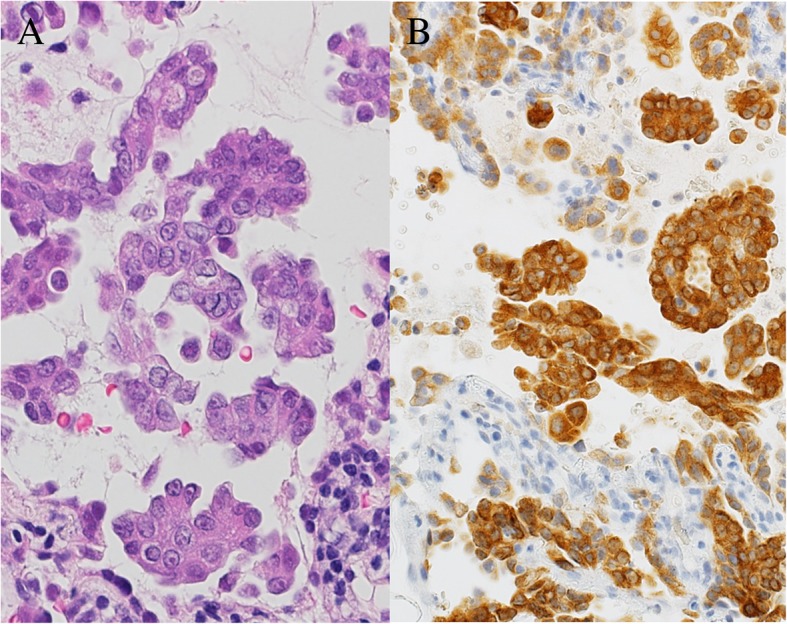
Hematoxylin and eosin stain of bronchoscopy for the middle lobe of the left lung, showing adenocarcinoma cells exhibiting a papillary pattern (a). Immunohistochemistry method for anaplastic lymphoma kinase from the same sample (b)

Hematoxylin and eosin stain of bronchoscopy for the middle lobe of the left lung, showing adenocarcinoma cells exhibiting a papillary pattern (a). Immunohistochemistry method for anaplastic lymphoma kinase from the same sample (b)