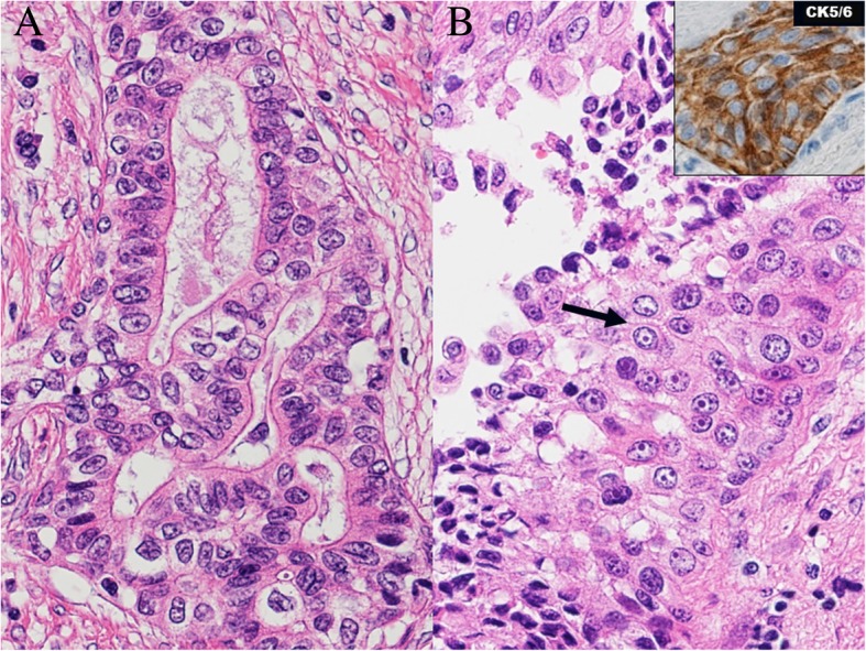
Fig. 4.

Hematoxylin and eosin stain of the primary lesion at necropsy. Approximately 20% of the lesion manifested several characteristics of squamous cell carcinoma as determined by the presence of intercellular bridges (arrow) and cytokeratin 5/6 positivity, indicating an adenosquamous carcinoma (a, b). CK5/6 cytokeratin 5/6
