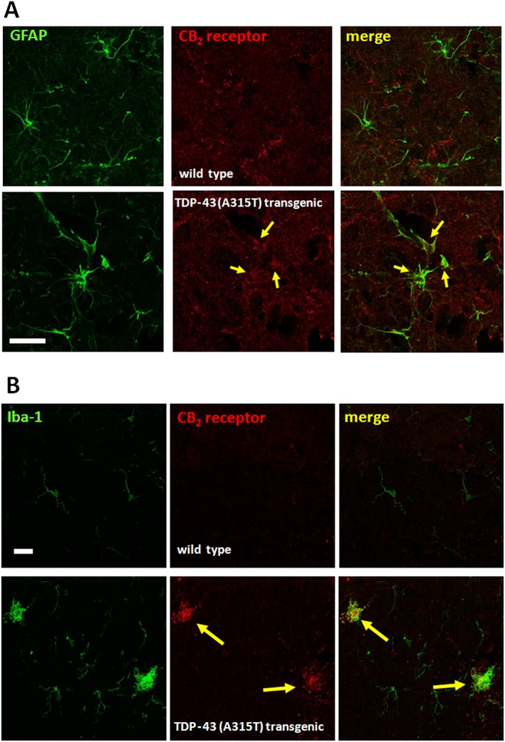
Figure 11.

Representative double‐immunofluorescence images for CB2 receptors and GFAP (dorsal horn; see panel A) or Iba‐1 (white matter; see panel B) in the spinal cord of TDP‐43 (A315T) transgenic and wild‐type male mice at 90 days after birth. Immunostainings were repeated in five animals per group. Cells positive for GFAP or Iba‐1 and CB2 receptors are indicated with arrows (scale bar = 25 μm).
