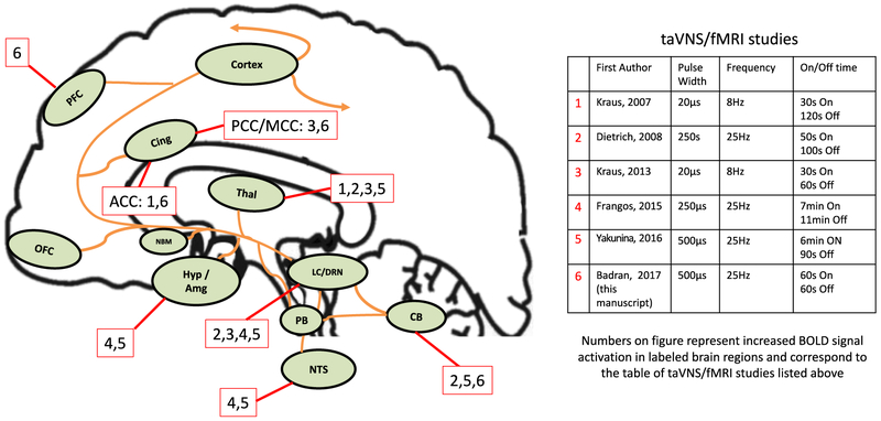
Fig. 1. Afferent pathway of the vagus nerve and regions activated by taVNS/fMRI studies.
Areas of the brain involved in the afferent vagal pathway. Nucleus tractus solitarius (NTS), locus coeruleus (LC), cerebellum (CB) thalamus (Thal), hypothalamus (Hyp), amygdala (Amg), and nucleus basalis (NBM) orbital frontal cortex (OFC), cingulate cortex (Cing), and prefrontal cortex (PFC). Effects are not limited to the named structures, as there are unlisted widespread, diffuse cortical effects (Cortex). Numbers labelling relevant regions of brain activations determined by corresponding study listed in adjacent table.

