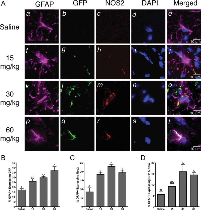
Fig. 4.
Neuroinflammatory activation of astrocytes occurs in NF-κB reporter mice prior to loss of dopaminergic neurons. A: Representative 40× images of astrocytes (GFAP; purple) expressing intrinsic GFP fluorescence (green), inducible nitric oxide synthase (NOS2; red), and counterstained with DAPI (blue). B: Quantification of the number of GFAP+astrocytes expressing intrinsic GFP. C: Quantification of the number of GFAP+astrocytes expressing NOS2. D: Quantification of the number of GFAP+astrocytes expressing both GFP and NOS2. Data are expressed as percentage ± SEM (n = 9). [Color figure can be viewed in the online issue, which is available at wileyonlinelibrary.com.]

