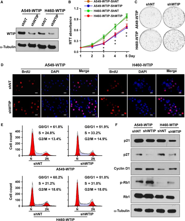
Figure 4.

Depletion of WTIP promotes cell proliferation and cell cycle progression. (A) Western blotting analysis of WTIP in cell lines with stable expression of specific shRNAs. (B) MTT assay analysis of cell growth. Error bars represent the mean ± SD obtained from three independent experiments. *P < 0.05, unpaired t‐test. (C) Representative pictures of cell colonies originating from the indicated cells and stained with crystal violet. (D) Representative pictures of BrdU staining. Pictures were taken at 400× magnification. Scale bars, 20 μm. (E) Cell cycle distribution as analyzed by flow cytometry. (F) Western blotting analyses of the expression of p21, p27, cyclin D1, phosphorylated Rb (p‐Rb), and total Rb in the indicated cells. α‐Tubulin served as a loading control.
