
Figure 4.
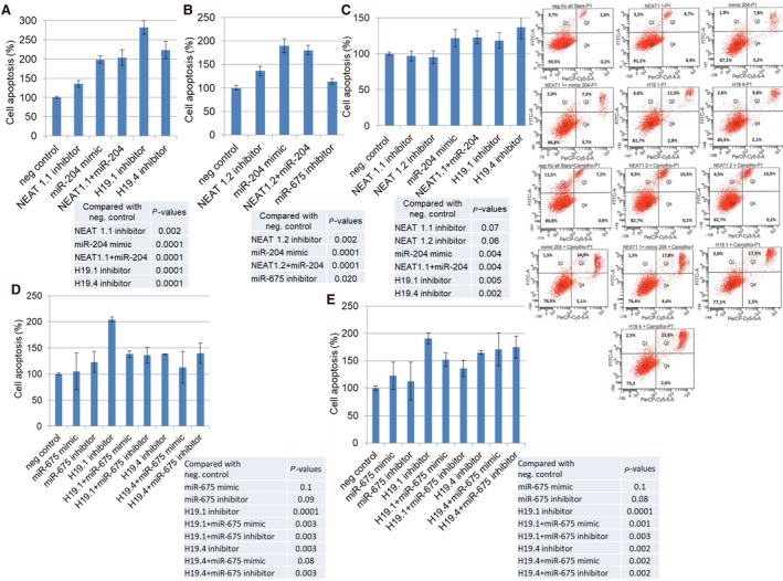
Effects on apoptosis by H19/miR‐675 and NEAT1/miR‐204. MCF cells were transfected with miScript inhibitor negative control, AllStars negative control small interfering RNA, NEAT1.1 inhibitor and/or miR‐204 mimic, and inhibitors of H19.1 and H19.4 (A), with miScript inhibitor negative control, AllStars negative control small interfering RNA, NEAT1.2 inhibitor and/or miR‐204 mimic, and miR‐675 (B), and additionally treated with the topoisomerase I inhibitor camptothecin (C), with miScript inhibitor negative control, AllStars negative control small interfering RNA, inhibitors of H19.1 or H19.4, miR‐675 mimic or inhibitor and both, H19.1 inhibitor plus miR‐675 mimic or inhibitor and H19.4 inhibitor plus miR‐675 mimic or inhibitor (D), and additionally treated with the topoisomerase I inhibitor camptothecin (E). The bar charts summarize the data on the apoptotic effect by these lncRNAs and miRNAs as derived from the FACSCanto II device. The experiments were repeated two times and showed similar results, indicated by error bars representing standard deviation. The P‐values refer to the comparison of the indicated ncRNAs with the average of the two neg. controls which displayed similar values. Below the bar charts, exemplary diagrams of the FACS analyses using MCF cells untreated and treated with Camptothecin are shown. Cells were labeled with Annexin‐V‐FITC and propidium iodide. Cell fragments only positive for propidium iodide can be found in the upper left corner (Q1). Late apoptotic as well as necrotic cells can be found in the upper right corner (Q2), since they are positive for Annexin and propidium iodide. Living cells are negative for Annexin and propidium iodide and, therefore, can be found in the lower left corner (Q3). Early apoptotic cells are only positive for Annexin and located in the lower right corner (Q4). The size for each population (%) is given in the corresponding area.