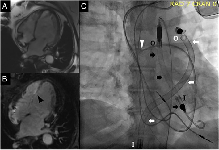
Figure 1.

(A) Four‐chamber view on cardiac magnetic resonance imaging showing thinning of entire inter‐ventricular septum; (B) delayed contrast‐enhanced four‐chamber view on cardiac magnetic resonance imaging showing fibrosis/scarring of the inter‐ventricular septum (black arrow head); (C) fluoroscopic image showing pulmonary artery catheter (white arrow head), Impella 5.0 (black arrows), and Impella RP (white arrows). The Impella 5.0 has its inlet in the left ventricle (black ‘I’) and outlet in the ascending aorta (black ‘O’), while the Impella RP has its inlet in the inferior vena cava–right atrial junction (white ‘I’) and outlet in the pulmonary artery (white ‘O’).
