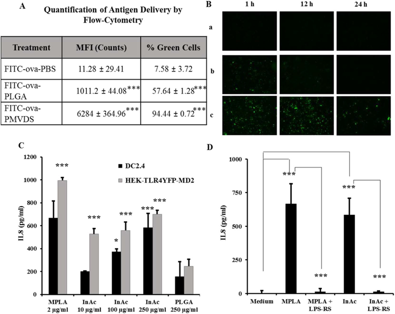
Fig. 4.
PMVDS targets dendritic cells for antigen delivery (A-B) and activation (C-D). (A-B) Mouse dendritic cells (DC2.4) were Incubated with the antigen, FITC-ova delivered through PBS (a), PLGA particles (b), or InAc-PMVDS (c) as a vehicle. (A) The amount of antigen delivered in 1 h was quantified by flow cytometer by measuring the mean fluorescence intensity (MFI) of each cell. Data represent mean ± standard deviation (n = 3) and *** represents that the values are significantly different when compared to FITC-ova-PBS group (at p < 0.001) using one-way ANOVA with Bonferroni’s multiple comparison test. (B) The persistence of the antigen delivered in 1 h to dendritic cells was followed at different time periods using fluorescence imaging (1 h, 12 h, and 24 h). (C-D) InAc particles activate dendritic cells through TLR-4. (C). Dendritic cells and HEK cells expressing only TLR-4 were incubated with a known TLR-4 agonist MPLA (2 μg/ml), InAc particles at different concentration (10, 100 and 250 μg/ml), and PLGA particles (250 μg/ml). Data represents mean ± standard deviation (n = 4–5). * and *** indicates that values are significant at p ≤ 0.05 and p ≤ 0.001 respectively, as compared to PLGA treated cells using one-way ANOVA followed by Dunnett’s post-hoc multiple comparison test. (D) Simultaneously, DC2.4 cells were incubated with an established TLR-4 antagonist, LPS-RS (120 ng/ml) for 1 h before adding InAc particles (250 μg/ml) or MPLA (2 μg/ml). After 48 h of immune stimulation, cell supernatants were collected and analyzed for the levels of IL-8 by ELISA as a readout for activation. InAc particles along with MPLA stimulated DC2.4 cells. However, LPS-RS inhibited InAc induced immune-stimulation of DC2.4 cells. Data represents mean ± standard deviation (n = 4–5). *** indicates that values are significant, at (p ≤ 0.001) using one-way ANOVA, followed by post-hoc multiple comparison tests. Dunnett’s multiple comparison test was used for MPLA and InAc group to compare with the medium treated cells. Bonferroni’s multiple comparison test was used for MPLA + LPS-RS and InAc + LPS-RS groups to compare with MPLA and InAc groups, respectively.

