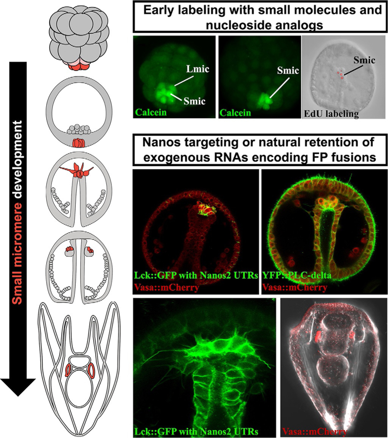
FIG. 1.

Illustrations of small micromere development from birth during cleavage stages through coelomic pouch residence in pluteus larva. Each stage of small micromere development can be labeled with small molecule dyes such as calcein-AM or nucleoside analogs like EdU. Later stages of small micromere development, including migration behaviors and left/right coelomic pouch distributions, can be observed after injecting RNAs encoding fluorescently tagged germline-specific genes or by engineering messages to contain the Nanos2 UTR retention sequences.
