
Fig. 3. HFCS treatment accelerates de novo fatty acid synthesis in intestinal tumors from APCdeficient mice.
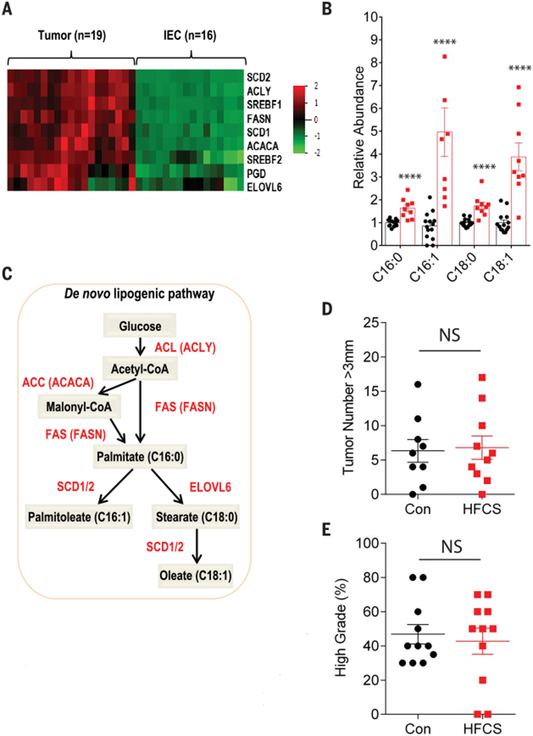
(A) Heatmap depicting the relative expression of the indicated genes involved in fatty acid synthesis from APC−/− tumors (n = 16) and intestinal epithelial cells (IECs, n = 16) using RNA-seq data. (B) Relative abundance of saturated and unsaturated 16- and 18-carbon fatty acid species in APC−/− tumors treated daily with water (Con, n = 14) or HFCS (n = 9). Groups compared by Student’s t test with correction for multiple comparisons using the Holm-Sidak method. ****P < 0.0001. (C) Schematic depicting key enzymes, genes, and metabolites in the de novo lipogenesis pathway. Red, enzyme name; red in parentheses, gene name. (D) APC−/−; FASN−/− mice were treated with a daily oral gavage containing water (Con, n = 9) or HFCS (n = 10) starting the day after tamoxifen injection and killed at 8 weeks. The size of each tumor (diameter) in the intestine was determined in whole-mount tissue after methylene blue staining, using a dissecting microscope. Data represent the number of tumors >3 mm in diameter in Con and HFCS-treated mice. Groups compared by Student’s t test. NS, not significant. (E) Percentage of high-grade tumors (n = 11 per group) from Con and HFCS-treated APC−/−; FASN−/− mice. Student’s t test. NS, not significant. All data represent means ± SEM.