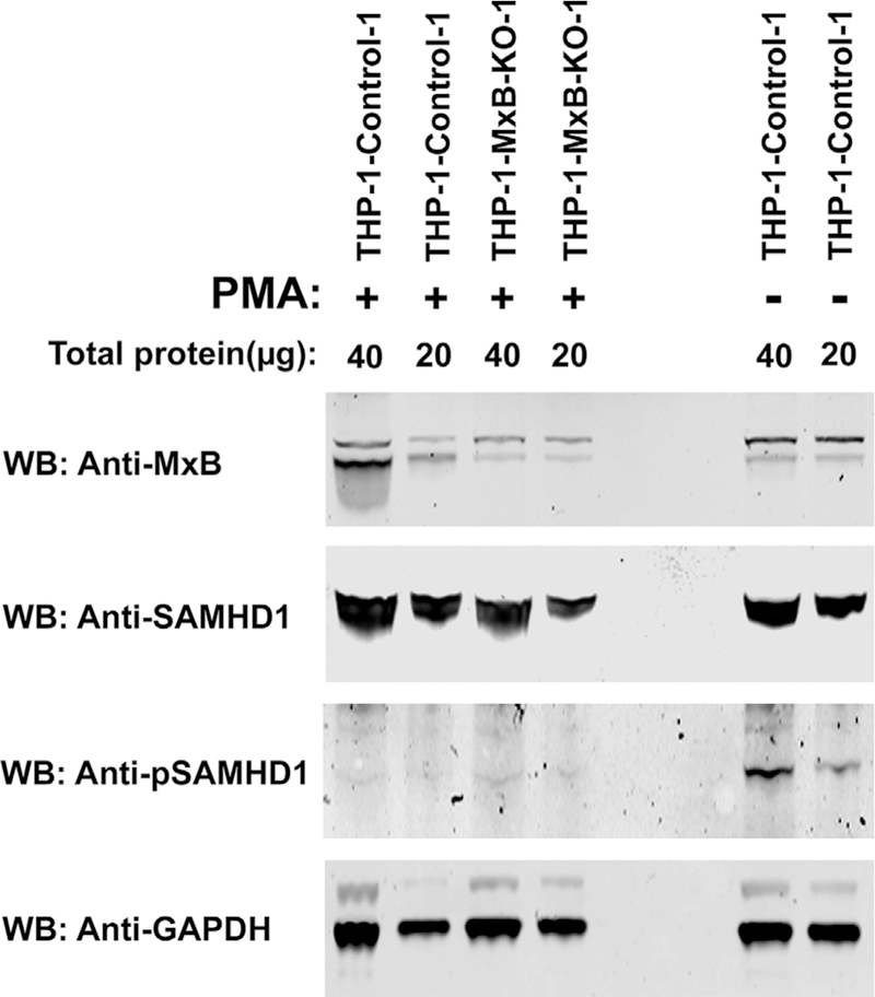
Fig. 9. Phosphorylation state of SAMHD1 at position T592 in the absence of MxB.

Lysates from PMA-treated THP-1 and THP-1-MxB-KO cells were analyzed by Western blot analysis using anti-MxB, anti-SAMHD1, and anti-phospho-T592-SAMHD1. As loading control, samples were analyzed for the expression of GAPDH. As a positive control for phosphorylation, THP-1 cells that were not treated with PMA were utilized. Experiments were done in triplicate and a representative experiment is shown.
