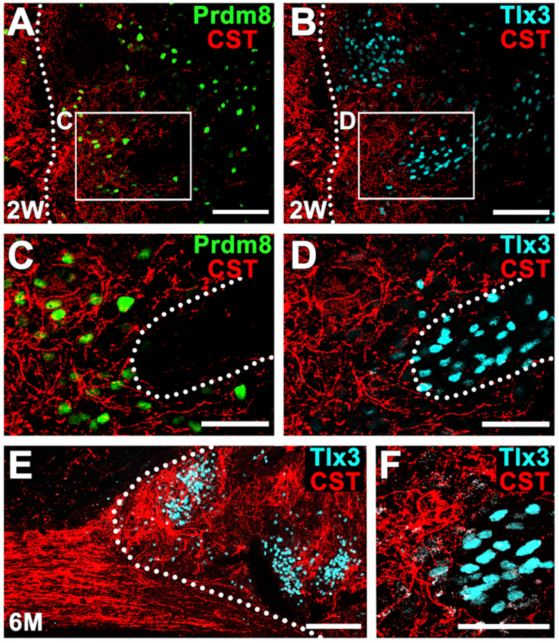
Figure 3. Host Corticospinal Axon Regeneration into Rat Neural Progenitor Cell Graft.

(A–D) Corticospinal axons (red) are present within regions of Prdm8-expressing V1-2 pre-motor interneurons (A and C) (green) and are attenuated in clusters of Tlx3-expressing sensory interneurons (B and D) (cyan) within the graft 2 weeks after grafting. Dotted lines indicate rostral host-graft border (A and B) and motor-sensory domain border (C and D). Sagittal section; rostral is left. Thin-plane confocal images of boxed areas are shown in (C) and (D). Scale bars, 100 μm (A and B) and 50 um (C and D).
(E and F) Six months after grafting, corticospinal axon density remains greater in grafts regions free of Tlx3-expressing sensory interneuronal clusters. Dotted line indicates host-graft border. Left, rostral; right, caudal. Scale bars, 200 μm (E) and 50 μm (F).
