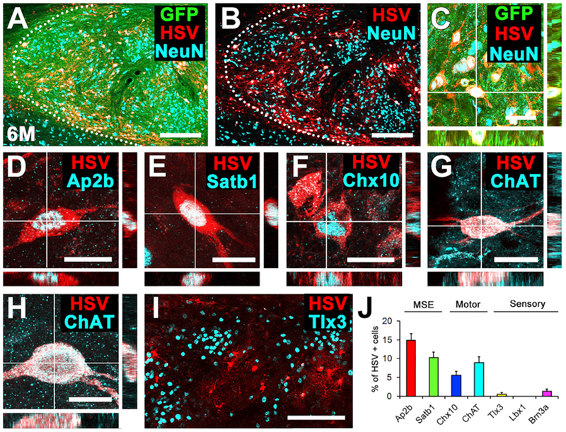
Figure 5. Anterograde Corticospinal HSV Tracing after 6 Months: Specificity of Graft Innervation from Host.

(A–C) Cells in grafts trans-synaptically labeled with tdTomato (red, HSV) are readily evident with the GFP-expressing graft, 6 months after implantation into spinal cord lesion cavity. Sagittal section; rostral left, caudal right. Dotted line indicates rostral host-graft border. Scale bars, 250 μm (A and B) and 50 μm (C).
(D–I) Characterization of graft neurons trans-synaptically labeled with tdTomato after motor cortex injections of HSV. Synaptically connected tdTomato+ graft-derived neurons include (D) Ap2b or (E) Satb1+ MSE neurons, (F) Chx10+ V2a, and (G and H) cholinergic neurons. Cholinergic neurons are likely to be both (G) V0c interneurons and (H) alpha motor neurons based upon their size and morphology. Scale bars, 20 μm.
(I) Trans-synaptically labeled tdTomato-expressing neurons located in domains outside inappropriate sensory interneuronal clusters (Tlx3+). Left, rostral; right, caudal. Scale bar, 200 μm.
(J) Quantification of HSV-tdTomato trans-synaptically labeled neurons (n = 4 animals). Means ± SEM.
