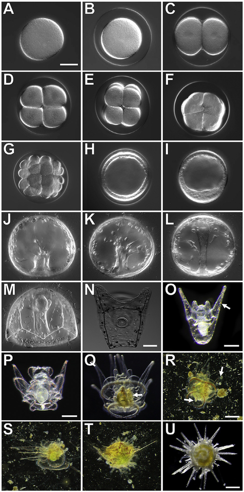
FIG. 4.
Developmental stages of the sea urchin, Lytechinus pictus. A–M and N imaged in DIC, scale bars = 50μM; O scale bar = 0.25mm; P and Q scale bar = 0.25mm; R–T scale bar = 0.25mm; U scale bar = 0.25mm. (A) Unfertilized egg; (B) Fertilized egg; (C) 2-cell embryo; (D) 4-cell embryo; (E) 8-cell embryo; (F) 16-cell embryo; (G) 32-cell embryo;(H) Early blastula stage; (I) Hatched mesenchyme blastula; (J) early gastrulation;(K) mid-gastrulation; (L) late gastrulation; (M) prism larva; (N) early 2-arm pluteus;(O) late 2-arm pluteus, red pigment cells (arrow) are visible in the ectoderm; (P) 8-arm pluteus without a rudiment; (Q) competent 8-arm pluteus with a rudiment (white arrow);(R) Early metamorphosis, a competent larvae swims to the substrate and tube feet (arrow) extend from the rudiment; (S) Mid-stage metamorphosis of a competent larvae, the animal moves the arms to the side and further attaches tube feet to the settlement surface, tissue along the arms is resorbed, revealing bare skeletal rods; (T) Late stage metamorphosis, the animal has extended the walking spines and the arms are now pushed to the top of the juvenile body as further tissue reorganization occurs; (U) Juvenile sea urchin at 1 week post-metamorphosis.

