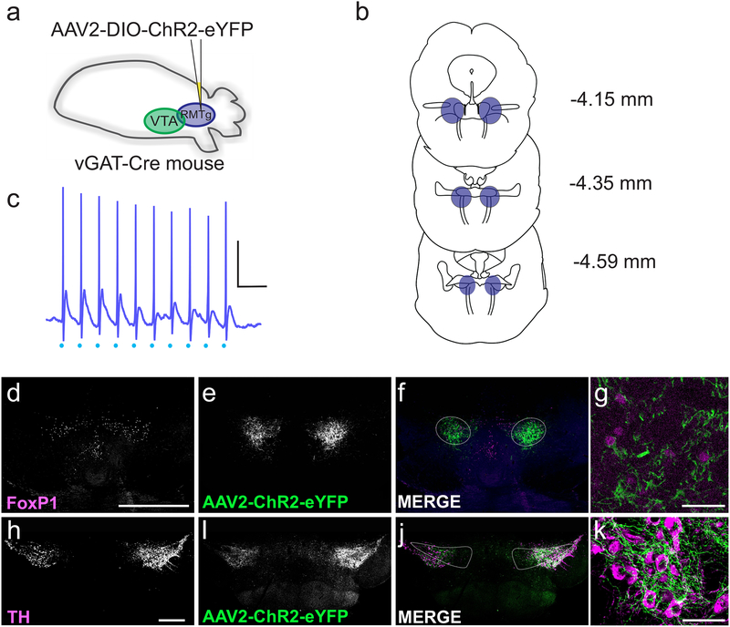
Figure 2. Isolating synaptic inputs from RMTg GABAergic neurons.
(a) Schematic: AAV2-DIO-ChR2-eYFP was injected into the RMTg (caudal to the VTA). (b) Coronal sections from 4.15 to 4.59 mm caudal to bregma illustrating the extent of the RMTg at each level; the region in blue represents the RMTg, targeted with injections. Coronal outlines adapted from Paxinos & Franklin, 2012. (c) Current clamp recording from a ChR2-expressing cell in the RMTg. Light stimulation (blue dots) at 20 Hz elicited action potentials in the cell; scale bar: 20 mV, 100 ms. (d-g) Example coronal section through the RMTg, demonstrating the expression of AAV2-ChR2-eYFP 3 weeks after viral injection. (d) FoxP1 staining identifies the RMTg; scale bar, 500 μm. (e) Expression pattern of AAV2-ChR2-eYFP. (f) Overlay of FoxP1 (magenta) and AAV2-ChR2-eYFP (green). (g) Co-labelling with AAV2-ChR2-eYFP (cytoplasmic; green) and FoxP1 (nuclear; magenta). Scale bar: 50 μm. (h-k) Example coronal section through the VTA, showing terminals from the RMTg expressing AAV2-ChR2-eYFP. (h) Immunohistochemistry for tyrosine hydroxylase (TH); scale bar, 500 μm. (i) AAV2-ChR2-eYFP in RMTg terminals within the VTA. (j) Overlay of TH (magenta) and AAV2-ChR2-eYFP (green). (k) RMTg fibers expressing AAV2-ChR2-eYFP (green) in close proximity to TH+ cell bodies (magenta). Scale bar: 50 μm.

