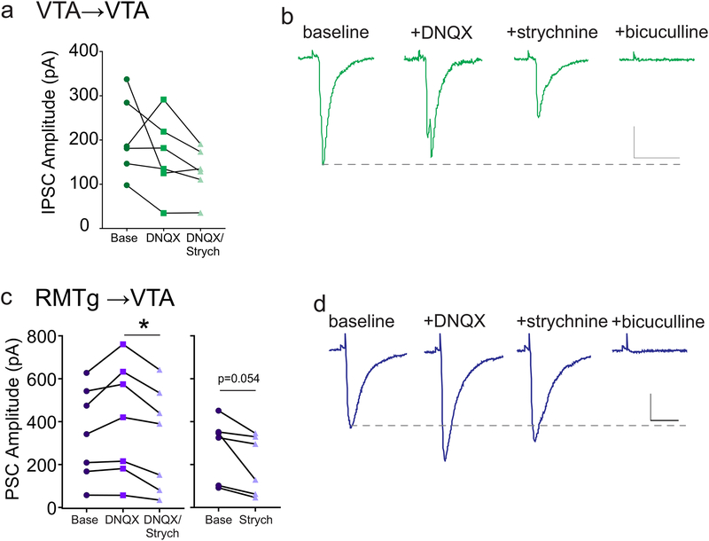
Figure 4. Neurotransmitter co-release from GABAergic terminals in the VTA and RMTg.
(a) Amplitude of light-evoked VTA→VTA PSCs were recorded before and after application of DNQX (10 μM), and then DNQX + strychnine (1 μM). (b) Representative PSCs during baseline, and during exposure to DNQX, DNQX + strychnine, and DNQX + strychnine + bicuculline (30 μM). No significant differences, 1-way ANOVA (n = 6 cells/6 mice). (c) Left panel: amplitudes of light-evoked RMTg→VTA PSCs recorded before and after application of DNQX (10 μM), and DNQX + strychnine (1 μM) (* p < 0.05, 1-way ANOVA; n = 7 cells/7 mice). Right panel, amplitudes of light-evoked RMTg→VTA PSCs recorded before and after application of strychnine alone (1 μM) (p = 0.05, paired t-test; n = 6 cells/5 mice). (d) Representative PSCs during baseline, and during exposure to DNQX, DNQX + strychnine, and DNQX + strychnine + bicuculline. Scale bars: 100 pA, 20 ms.

