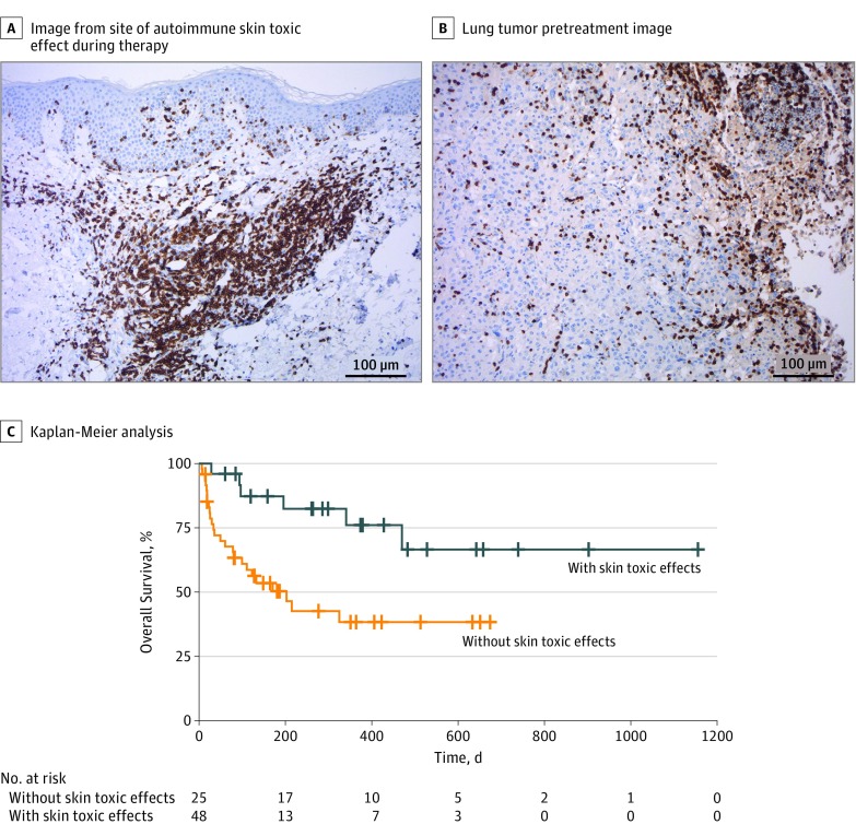
Figure 2. Association Between Autoimmune Skin Toxic Effects and Response to Therapy .
Images of a representative patient show autoimmune skin toxic effects (A) and lung tumors (B) characterized by an inflammatory infiltrate shown by CD3 immunohistochemistry (magnification ×100). C, Kaplan-Meier analysis of patients treated with PD-1 inhibitors reveals better outcome for 25 patients who developed skin toxic effects (blue line) compared with 48 other patients who did not develop skin toxic effects (orange line). One-year overall survival rate was 76% in the group with skin toxic effects and 38% in the group without skin toxic effects, with a hazard ratio of 0.29 (95% CI, 0.12-0.71) and log-rank P = .004.

