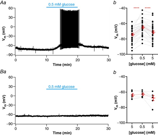
Figure 3. Most NTS neurons depolarize in response to a low glucose challenge.

A, subset of neurons depolarized by low glucose, as shown by representative recording of a responsive neuron showing a reversible depolarization and increased firing activity induced by low glucose (Aa) and summary of the low glucose effect on the membrane potential (V m) of neurons (Ab). B, subset of neurons unresponsive to low glucose, as shown by a representative recording (Ba) and summary of the low glucose effect on the V m of neurons (Bb). **** P < 0.0001. [Color figure can be viewed at wileyonlinelibrary.com]
