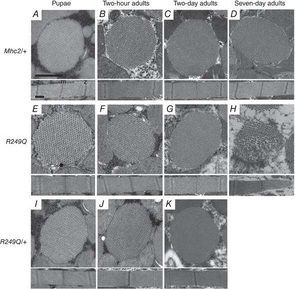
Figure 3. Transmission electron micrographs of R249Q IFM cross‐sections (top) and longitudinal sections (bottom).

Four different stages of development were analysed: late stage pupae, 2‐h‐old adults, 2‐day‐old adults, and 7‐day‐old adults. Mhc2/+ controls at all ages show that thick and thin filaments are arranged in a normal double‐hexagonal pattern and that myofibril morphology remains intact (A–D). We have shown previously that pwMhc2 and wild‐type fibres show no differences in myofibril and sarcomere formation (Swank et al. 2000). Sarcomere structure with regularly spaced M‐ and Z‐lines are evident at all stages. Homozygous R249Q late stage pupae and 2‐h‐old adults are essentially identical to Mhc2/+ (E and F). Homozygous R249Q fibres begin to experience a very small amount of disruption in fibril morphology and hexagonal packing at the edges of the myofibril at 2 days old (G), but disruption is evident at 7 days old (H). Heterozygous R249Q late stage pupae, 2‐h‐old adults, and 2‐day‐old adults are essentially identical to Mhc2/+ (I–K). Myofibrils shown in each panel are representative of the entire population at that given stage of development. Scale bars, 0.5 μm. M line and Z disk are labelled as ‘m’ and ‘z’, respectively, in panel A.
