Figure 4.

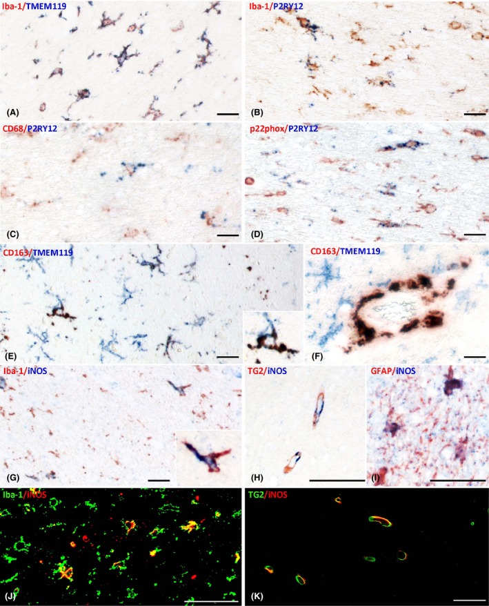
Patterns of microglia activation shown by double staining. (A–D) Coexpression of different markers in the white matter of a patients with sepsis shows that nearly all Iba‐1 positive cells also express TMEM119 (A), but only a fraction is P2RY12 positive (B); despite the expression of the ‘homeostatic’ marker P2RY12 some microglia also express the activation markers CD68 (C) of p22phox (D). (E–F) When CD163 is present in cells with microglia morphology they are also stained for TMEM119 (E and insert); in contrast, perivascular macrophages are CD163+ but TMEM119 negative (F). (G–K) iNOS is expressed in microglia (G,J) and astrocytes (I) in addition in endothelial cells in sepsis as well as in controls (H,K). Magnification Bars: 50 μm.
