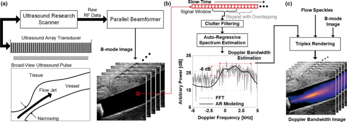
Figure 1.

Conceptual illustration of DUBI. (a) Plane wave data acquisition at a region of interest using a research‐purpose ultrasound scanner that generates high‐frame‐rate ultrasound images; (b) Overview of the Doppler bandwidth estimator for a slow‐time ensemble extracted from the B‐mode images (indicated by the red box in (a)), with the estimated Doppler power spectrum (dashed line) and the AR estimated spectrum (solid line) shown for illustration; (c) Doppler bandwidth images (DUBI frames) generated by overlaying color‐coded Doppler bandwidth estimates on top of flow speckles and the B‐mode image. [Color figure can be viewed at wileyonlinelibrary.com]
