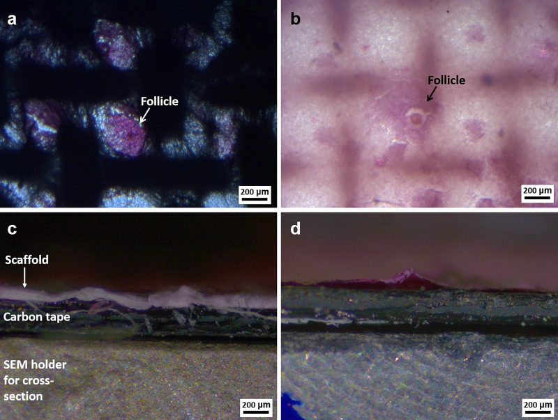
Fig 3.
Light microscopy images related to the top view and the cross-section of the patterned electrospun scaffolds seeded with porcine ovarian follicles 10 days after the seeding: (a,c) PCL and (b,d) blend of PCL and gelatin. An indication of the scaffold, carbon tape and holder is reported in c. For all images: magnification 5X, scale bar 200 μm.

