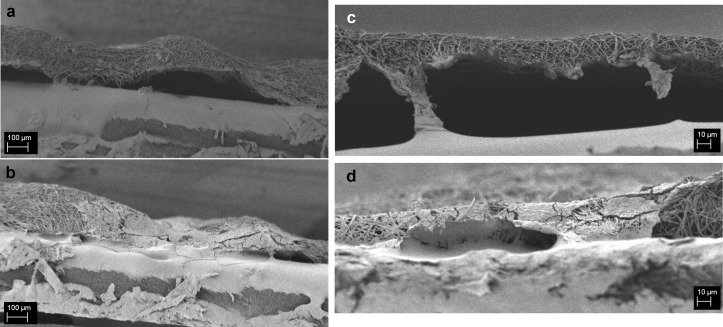
Fig 4.
SEM micrographs (a,b) of PCL and (c,d) PCL/gelatin scaffolds without follicles (a,c) and with follicles (b,d) 10 days after the seeding (H&E staining). For PCL scaffolds, the magnification is 150X and the scale bar is 100 μm for PCL/gelatin scaffolds the magnification is 1000X and the scale bar is 10 μm.

