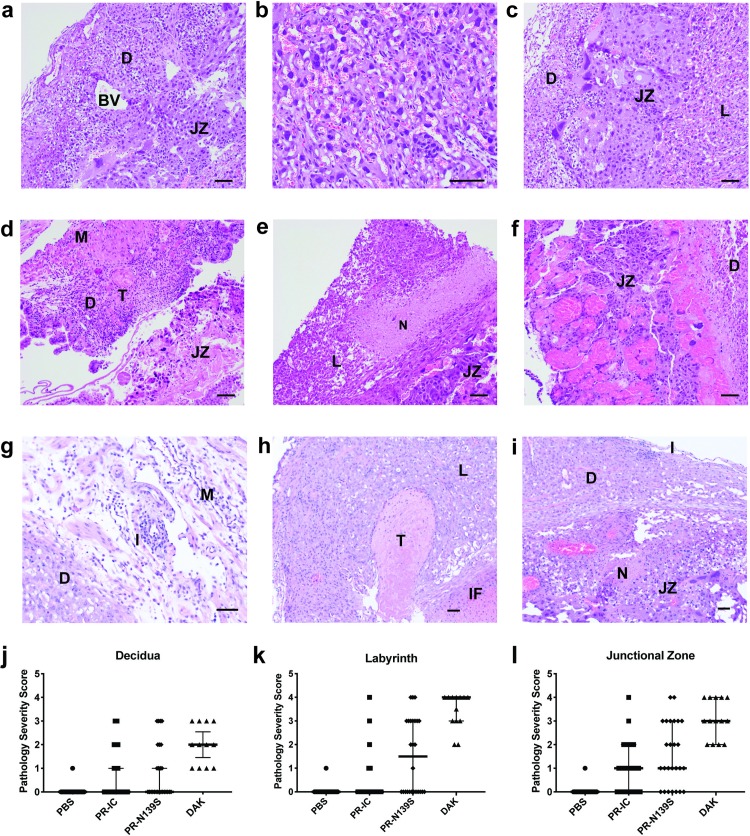
Fig 3. Placenta histopathology analysis: Hematoxylin and eosin (H&E) staining of placenta and fetus.
(a-c) Normal histologic features of each placental zone (decidual layer (D), labyrinth layer (L), and junctional zone (JZ)) from concepti from dams inoculated with PBS. BV, normal decidual blood vessels. (d-f) Severe histopathologic injury patterns for each zone from placenta from ZIKV-inoculated dams. (d) Myometrium (M) and decidua (D) from a ZIKV-PR-IC placenta with increased inflammation, multiple thrombi (T) in the decidua, and a necrotic JZ. (e) L from a ZIKV-DAK placenta with focal necrosis (N), lack of blood in most vascular spaces, and numerous degenerating cells. (f) JZ from a ZIKV-DAK placenta with markedly dilated blood vessels, focal thrombi, and a layer of necrosis at the interface with the decidua. (g) D and M from a ZIKV-PR-N139S placenta with inflammation (I). (h) L from a ZIKV-PR-N139S placenta with T and infarction (IF). (i) JZ from a ZIKV-PR-IC with N and I. (j-l) The degree of placental pathology was rated on a scale of 0–4: zero represents normal histologic features and 4 represents the most severe features observed. Each zone of the placenta was scored individually for general overall pathology, amount of inflammation, and amount of vascular injury with a consensus score for each placenta derived from three independent pathologists. Only ‘General’ scores are shown because they were representative of the ‘inflammation’ and ‘vascular injury’ categories and do not differ significantly from ‘general’. Error bars represent 95% confidence interval from the median. Data are representative of 3–5 independent experiments for each treatment group. Scale bar, 50 μm.

