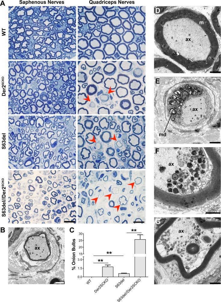
Fig 7. Morphology of Der2SCKO and S63del//Der2SCKO adult quadriceps and saphenous nerves.
(A) Transverse semithin sections of saphenous and quadriceps nerves from WT, Der2SCKO, S63del and S63del//Der2SCKO at 12 mo. Red arrowheads indicate onion bulbs. n = 4–5 nerves/genotype. Scale bar, 10μm; 100x magnification. (B) Image of an onion bulb by EM analysis on 12 mo S63del//Der2SCKO quadriceps nerves. Scale bar, 2μm; ax, axon. (C) Number of onion bulbs in 12 mo quadriceps nerve (expressed as a percentage of total myelinated fibers in the whole nerve); n = 3–4 nerves/genotype. Error bars, SEM; *P < 0,05, **P < 0,01 by Student’s t test. (D-G) EM analysis on 12 mo quadriceps and saphenous nerves. In (D), a normal fiber from WT quadriceps nerve is shown as control. Panel (E) shows a demyelinating fiber from S63del//Der2SCKO quadriceps nerves in which the cell surrounding the axon is full of myelin debris (md). In (F) and (G), signs of axonal degeneration detected in S63del//Der2SCKO quadriceps and saphenous nerves, respectively. (D-E) scale bar, 2μm; (F-G), scale bar, 1μm.

