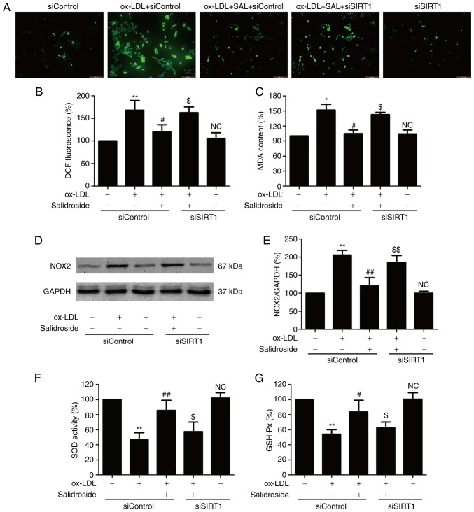
Figure 3.
Effects of SIRT1 knockdown on the inhibition of salidroside on ox-LDL-induced oxidative stress in HUVECs. HUVECs were pre-transfected with siSRT1 or siControl and incubated with SAL (10 µM) for 1 h followed by treatment with ox-LDL (100 µg/ml) for 24 h. (A) The intracellular ROS production was determined by a redox-sensitive fluorescent probe DCFH-DA staining. Scale bar, 200 µM. (B) The fluorescence intensity of DCFDA-stained sample was quantified by flow cytometry. (C) The intracellular MDA content was measured using the thiobarbituric acid method with a maximum absorbance at 532 nm. (D) The expression of NOX2 was determined using western blot analysis. (E) Bar charts demonstrate the quantification of NOX2 expression. (F) The SOD activity was detected using a colorimetric assay kit based on the combination of xanthine and xanthine oxidase. (G) The GSH-Px level was determined using a colorimetric assay kit depending on the enzyme-catalyzed reaction product (reduced glutathione). Data are expressed as the mean ± standard deviation; n=3, *P<0.05 and **P<0.01 vs. the control group; #P<0.05 and ##P<0.01 vs. the ox-LDL treatment group; $P<0.05 and $$P<0.01 vs. the ox-LDL and SAL co-treatment group. HUVECs, human umbilical vascular endothelial cells; ox-LDL, oxidized-low density lipoprotein; GSH-Px, glutathione peroxidase; SOD, superoxide dismutase; DCFH-DA, dichloro-dihydro-fluorescein diacetate; NOX2, NADPH oxidase 2; MDA, methane dicarboxylic aldehyde; ROS, reactive oxygen species; SAL, salidroside; NC, not significant; si, small interfering.

