Figure 1.
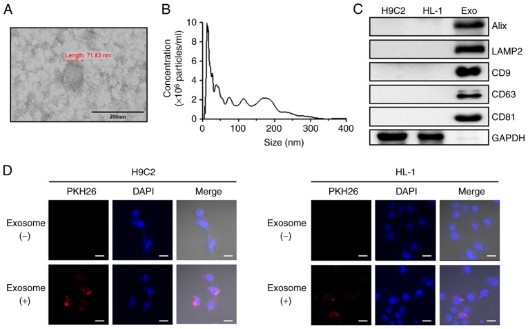
Characterization of exosomes isolated from human peripheral blood and uptake of exosomes by cardiac cells. (A) Morphology of human peripheral blood-derived exosomes imaged using transmission electron microscopy (scale bar, 200 nm). (B) Human peripheral blood particle size/concentration were analyzed by nanoparticle tracking analysis. (C) Protein expression were determined using western blot analysis using the indicated exosome marker antibodies. (D) H9C2 and HL-1 cells were treated with unlabeled or PKH26 (red)-labeled exosomes for 24 h and analyzed by confocal microscopy (scale bar, 20 µm; magnification, ×800). Cell nuclei were stained with DAPI (blue). Alix, programmed cell death 6 interacting protein; LAMP2, lysosomal associated membrane protein 2; CD, cluster of differentiation.

