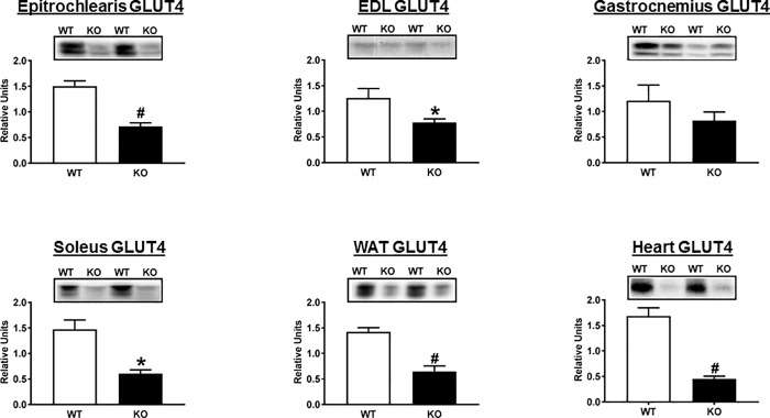
Fig 6. Total abundance of GLUT4 in tissues collected immediately after the hyperinsulinemic-euglycemic clamp performed in WT (open bars) and KO (filled bars) rats.
EDL, extensor digitorum longus; WAT, white adipose tissue. Bar graphs represent the ratio of the values for the immunoblots and their respective loading controls (Memcode stain). *P<0.05 and #P<0.001 for WT versus KO rats. Values are means ± SEM. N = 5–6 rats per group.

