
Figure 3:
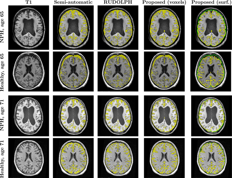
Comparison of semi-automatic and automatic methods, showing an axial slice of the subject’s T1, semi-automatic method result, RUDOLPH result, the proposed method after conversion to a binary map on a voxel grid, and proposed method as an inner (yellow) and outer (green) subarachnoid surface.