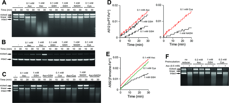
Figure 5. Plasmid DNA breakage by BLM in the presence of different reducers.
Reactions were performed at 37°C and contained 25 mM HEPES, pH 7.4, 0.5 μg ϕX174 plasmid DNA, 0.1 μM BLM and 0.1 μM Fe(III) citrate. (A) Plasmid DNA breakage by BLM-iron(III) in the presence of different reducers. (B) DNA breakage by Fe(III) citrate in the absence of BLM. (C) DNA breakage by BLM-Fe(III) in the mixtures of Asc with other reducers. (D) Fe(II) formation measured by the o-phenanthroline (o-PT) assay in the reaction of Fe(III) citrate with cellular reducers (37°C reactions in 25 mM HEPES, pH 7.4). Shown are means of triplicate measurements. (E) Ferrozine assay for Fe(II) formation in the reaction of Fe(III) citrate with cellular reducers. Shown are means of triplicate measurements. (F) Self-inactivation of BLM in the absence of DNA. BLM-Fe(III) was preincubated with the indicated reducers for 5 min at 37°C prior to the addition of plasmid DNA and 0.5 mM Asc. The final mixtures were incubated for 10 min at 37°C.

