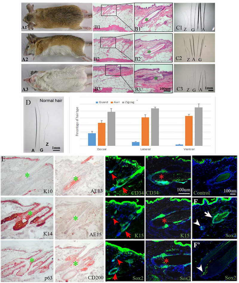
Fig. 1.
Spiny mouse hair types. A1. Dorsal view of adult spiny mouse with dark brown spiny hairs. A2. Lateral view of adult spiny mouse with yellow spiny coat. A3. Ventral view of adult spiny mouse with white spiny coat. B1, 2, and 3. H&E staining of dorsal, lateral, ventral spiny mouse skin. B1’, B2’, B3’, the magnified view of B1, B2, and B3, respectively. C1, C2, and C3, all hair types (Z: zigzag hair; G: guard hair; A: awl hair) from dorsal, lateral and ventral side of the skin, respectively. D. The percentage of hair types from 1 cm2 area of each respective body part. SD is calculated from 3 independent samples (N=3); more than 400 hairs are calculated in each sample. E. Immunostaining of K10, K14, p63, AE13, AE15, CD200, CD34, K15, and Sox2. Control: secondary antibody negative control. Arrowhead: zigzag hair; arrow: guard hair; asterisk: awl hair. F. Sox2 immunostaining of telogen and anagen hair follicles. White arrow: hair bulge, arrowhead: dermal papilla. All photographs are representative of the data acquired from all specimens.

