Figure 2.

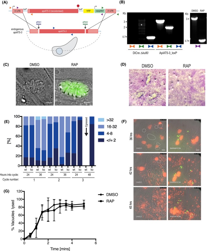
ApiAT5‐3 is essential for parasite proliferation. A. Generation of the ApiAT5‐3_loxP line using CRIPSPR/Cas9 to increase site‐directed integration. Protospacer adjacent motif (PAM) indicated by black arrows. Primer pairs represented by coloured triangles. B. Left panel: PCR analysis shows correct integration of the ApiAT5‐3_loxP construct at both the 3ʹ and 5ʹ ends and a loss of WT apiAT5‐3 at the endogenous locus. White * = non‐specific bands. Right panel: Addition of RAP leads to correct recombination of the loxP sites. C. Fluorescent microscopy of ApiAT5‐3_loxP parasites 24 h after addition of DMSO or RAP. Scale bar 5 µm. D. Plaque assay showing loss of plaquing capacity of ApiAT5‐3_loxP parasites upon RAP treatment. E. Parasite per vacuole number shown as mean %, n = 3. F. Stills from live video microscopy at 36, 42 and 45 h into third lytic cycle post‐RAP treatment. Red = RH Tom, dashed white line = intact WT ApiAT5‐3_loxP vacuoles, green = apiAT5‐3 KO. Scale bar 20 µm. G. IIE assay showing no significant difference between DMSO‐ and RAP‐treated ApiAT5‐3_loxP (at 30 h into lytic cycle 2 post‐DMSO/RAP treatment). Statistical analysis using multiple comparison two‐way ANOVA, n = 2.
