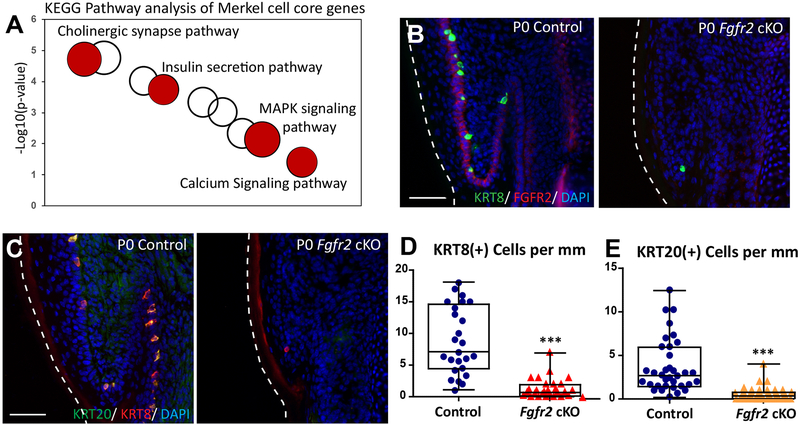
Figure 4. FGFR2 is critical for Merkel cell formation in the glabrous paw skin.
(A). KEGG pathway analysis of Merkel cell core genes from the back skin and glabrous paw skin. Selected KEGG terms are in red. The size of a cycle indicates the number of genes in a KEGG term. (B). Immunofluorescence analysis of Merkel cell markers KRT8 (green) and FGFR2 (red) in P0 control and Fgfr2 cKO glabrous paw skin. Note that FGFR2 (red) is completely gone in P0 Fgfr2 cKO. (C). Immunofluorescence analysis of Merkel cell markers KRT20 (green) and KRT8 (red) in P0 control and P0 Fgfr2 cKO glabrous paw skin. (D). Quantification of KRT8 (+) Merkel cells in P0 control and P0 Fgfr2 cKO, p < 0.0001, n = 4. (E). Quantification of KRT20(+) Merkel cells in P0 control and P0 Fgfr2 cKO, p < 0.0001, n = 4. White dotted lines indicate the edges of the tissue. Scale = 50μm in (B) and (C). The data presented in box plots (D, E) show the median with 25th and 75th percentile borders. Whiskers extend from minimum to maximum. ***p < 0.001 (per Mann-Whitney test).

