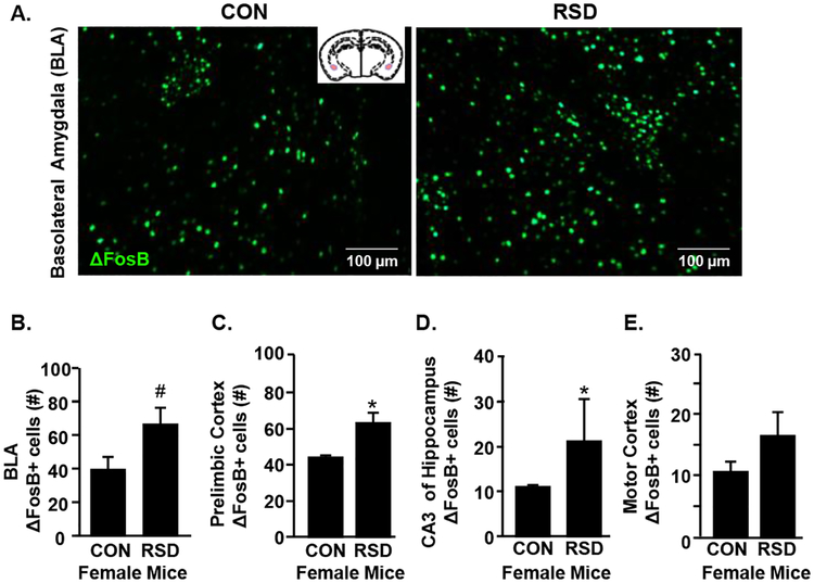
Figure 2. RSD in Female Mice Activates Neurons in Brain Regions Associated with Fear and Threat Appraisal.
Adult female C57BL/6 mice were subjected to six cycles of modified repeated social defeat (RSD) for 30 minutes per day using a male DREADD aggressor. Mice were perfused and PFA-fixed 14 h following the final cycle of RSD. Brains were sectioned and ΔFosB was determined in the Prelimbic Cortex, Basolateral Amygdala (BLA), CA3 of the hippocampus, and the Motor Cortex (n=6 per group). A) Representative images of ΔFosB labeling in the BLAof control and RSD mice. Number of ΔFosB+ cells in the B) BLA, C) prelimbic cortex, D) CA3 region of the hippocampus, and E) motor cortex. Bars represent the mean +/− SEM. Means with asterisks (*) are significantly different than controls (p <0.05). Means with (#) showed a trend (p = 0.06) to be different from controls.

