
FIG. 3.
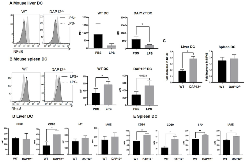
DAP12 negatively regulates NFκB and cell surface costimulatory molecule expression by liver DC. (A, B) WT or and DAP12−/− B6 mice were injected with 100ng E.coli LPS or PBS (control) via the portal vein and euthanized 2 hours later. (A, B) NFκB expression in freshly-isolated liver DC (A) and spleen DC (B) was assessed by flow cytometry. Left-hand histograms show representative flow data. Right hand panels show aggregate data from n=3 independent experiments. Compared with PBS injection, NFκB expression was reduced in liver DC 2 hours after LPS injection, but increased in splenic DC. (C) Whereas the fold increase in NFκB in spleen DC was similar for WT and DAP12−/− spleen DC after LPS injection, there was significantly less reduction in DAP12−/− liver DC compared with WT liver DC. Data are means + SEM obtained from n = 3 independent experiments. (D, E) Expression of MHC class II (IAb) and costimulatory molecules by WT and DAP12−/− liver DC (D) and spleen DC (E) (following LPS stimulation for 2 hours in vivo). *p<0.05; **p<0.001.