
Figure 4:
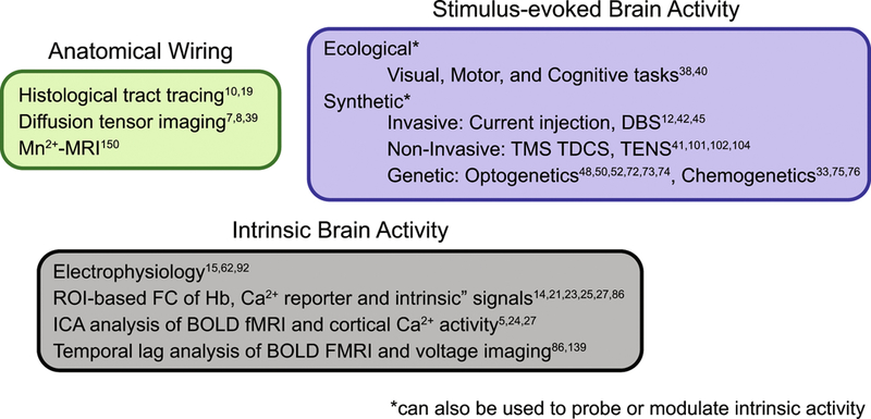
Tools for mapping brain connectomics. Mapping anatomical, or white matter, connections of the brain can be performed with ex vivo histological tract tracing(10, 19), or in vivo using diffusion tensor imaging (7, 8, 39) or manganese-enhanced MRI(150). Mapping the functional organization of the brain can be performed in vivo by invoking ecological (physiologically normal) stimuli as functional localizers (38, 40), or via synthetic (non-physiological) stimuli via invasive (12, 42, 45), non-invasive(41, 101, 102, 104), or genetics-based (33, 48, 50, 52, 72–76) strategies. In the absence of any overt task, ongoing spontaneous or intrinsic brain activity can be evaluated through electrophysiology(15, 62, 92), ROI-based (14, 21, 23, 25, 27, 86) or ICA-based(5, 24, 27) analyses, as well as through evaluating the relative timing differences in regional activity propagation (temporal lag analysis) (86, 139). Abbreviations: Mn2+-MRI: manganese enhanced magnetic resonance imaging; DBS: deep brain stimulation; TMS: transcranial magnetic stimulation; TDCS: transcranial direct current stimulation; TENS: transcutaneous electrical nerve stimulation; ROI: region of interest; Ca2+: Calcium; ICA independent component analysis; BOLD: Blood oxygen level dependent; FMRI: Functional magnetic resonance imaging.