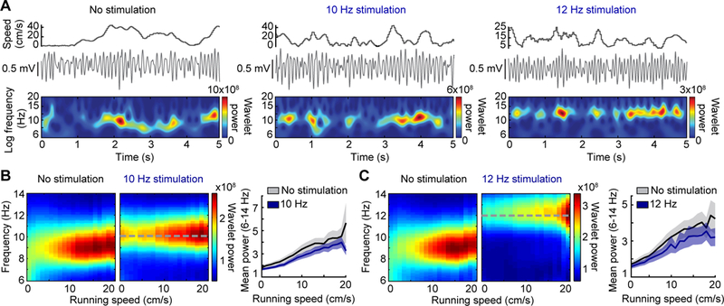
Figure 2. Theta amplitude increases with running speed during pacing.

(A) Hippocampal LFP and wavelet power is displayed as a function of running speed for periods without stimulation, 10 Hz stimulation, and 12 Hz stimulation of septal PV neurons. Note that the wavelet power was highest during periods with fast running speeds in both baseline and stimulation conditions. (B, C) (Left) Mean spectrograms over all recording sessions show the amplitude and frequency distribution across running speeds. (Right) Mean (± SEM) normalized power of theta oscillations (averaged over 6–14 Hz) is plotted by running speed. Stimulation controlled the frequency but did not preclude the modulation of theta amplitude by running speed during either 10 Hz or 12 Hz stimulation.
