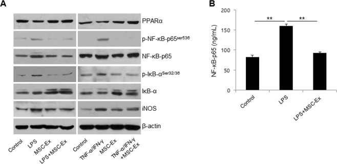
Figure 2.
MSC-Ex restores the PPARα activity in HaCaT cells. (A) HaCaT cells were treated as described in Fig. 1 and western blot analyses were performed using primary antibodies for PPARα, NF-κB-65, p-NF-κB-p65Ser536, IκB, p-IκBSer32/36 and iNOS. β-Actin was used as the loading control. (B) The protein levels of NF-κB were determined by ELISA. Columns represent the means of three independent experiments; bars indicate standard deviations.). *P < 0.05, **P < 0.005, ***P < 0.001.

